Leveraging 3D Imaging Data for Accurate Treatment Planning
It’s been a decades-long journey for one patient and his No. 8 tooth, beginning with trauma to the tooth at age 18 and continuing with incomplete endodontic treatment years later, but it will finally be resolved thanks to the data provided by 3D imaging.

The 45-year-old male patient initially presented to our office in May 2015 with the chief complaint of a hole in the back of No. 8 (Figure 1). Periapical radiographs were taken and a possible apical root fracture was observed based on certain x-rays (Figure 2). The patient had endodontic treatment started on the tooth in 2009, but had never returned to complete the treatment. Eventually, the temporary restoration dislodged from the lingual, prompting the patient to visit our dental practice in 2015.
Cotton was present in the canal, and a gingival abscess was traced to the mid-level of the tooth radiographically (Figure 3). At this point, we completed the endodontic procedure for No. 8 up to the point where we believed the root to end (Figure 4), noting that there was a possible fracture further apical to that point. A lingual composite was placed on the tooth, and the patient was made aware that the prognosis for the tooth was guarded. However, he did not return for six years, and in February 2022, the patient visited the office with the chief complaints: “My front tooth is painful, it moves, it is discolored, and sometimes it has an abscess.”
The examination revealed that tooth No. 8 had Grade 3 mobility, with suppuration emanating from the sulcus upon palpation of the gingiva. The periapical radiograph showed resorption of the root at the point where the gutta percha fill of the root canal treatment ended (Figure 5). The apical fracture of No. 8 was more clearly noted on this image. The apical portion seemed ankylosed. My initial assessment based on the 2D images was to extract and evaluate for a bone graft/membrane placement, allow 3-4 months of healing time, and then reassess for implant placement. The patient inquired if there was any chance for the implant to be placed at the time of extraction and whether the fractured part of the root would be removed.
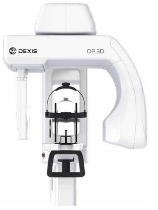
A CBCT scan was taken using the DEXIS ORTHOPANTOMOGRAPH™ OP 3D™ x-ray imaging system to determine the appropriate treatment plan. We utilized the endodontic resolution protocol (95 kVp, 2 mA, 80 µm). The patient also had teeth on the lower arch that were compromised and wanted both arches scanned, as implants on the lower arch are possible treatment avenues that the patient may want to explore.
Treatment Plan
With the ability to view the patient’s tooth No. 8 in 3D, and with the aid of DTX Studio Clinic software visualization, we were able to make several definitive diagnoses (Figure 6). We discovered a small root fragment at the alveolar crest on the palatal aspect (Figure 7). The apical fracture was sizable, but it did not appear to be as ankylosed as I originally hypothesized. The suppuration appeared to emanate from the mid-root area, with no visible pathology around the apical fracture of the root (Figure 8). Finally, the quality and quantity of bone was found to be viable for dental implant placement.
If we are successful in removing the root tip in atraumatic fashion and debride the area of all infectious tissue, then we may be able to place an immediate implant at the No. 8 site. We would create a temporary using the patient’s actual tooth No. 8 by removing the root from it and then applying a lingual splint that bonds the coronal No. 8 to the adjacent teeth (the patient has a Class III bite). We would allow 3-4 months for osseointegration and then restore No. 8 accordingly.
Conclusion
3D imaging data provides a level of clarity above and beyond the limitations of 2D intraoral and extraoral imaging. In this clinical case, although the apical fracture of No. 8 was seen on the radiographs, the OP 3D CBCT scan data revealed another path toward restoring the patient’s failing tooth. We were able to visualize the true size of the apical fracture for No. 8 as well as the full dimensions of the bone.
Performing an immediate implant placement at the site of No. 8 will shorten the amount of time that the patient will need to wear a temporary tooth by months, which is something any patient would appreciate. The patient was initially unsure about how to proceed, but after I reviewed the scan with him using "Endo" 3D visualization in DTX Studio Clinic (Figure 9) and discussed the possibility of immediate implant placement, the patient agreed with the proposed treatment plan. As the case shows, CBCT imaging not only enables accurate treatment planning, but greater case acceptance.

OP 3D
The OP 3D makes choosing your x-ray system simple. It is a complete x-ray platform that provides easy-to-use features throughout the entire dental imagine workflow. With its versatile imaging programs and intuitive user interface, the OP 3D in its different configurations offers imaging excellence for a variety of users, ranging from general dental practitioners to orthodontists, and all the way to maxillofacial surgeons. The OP 3D is designed to be upgradeable, allowing it to grow with the needs of your practice. The cephalometric or 3D imaging capabilities can be added at any time.

After graduating from the New Jersey Dental School at the University of Medicine and Dentistry at New Jersey, Dr. Gaur completed a General Dental Residency program at Jamaica Hospital Medical Center in Jamaica, NY. He then worked as a dentist in a private practice in the Triboro area of New York before purchasing his own private practice in 2012 on Long Island, NY. When not caring for patients at his general and pediatric dentistry office, Dr. Gaur enjoys maintaining an active lifestyle, traveling, rooting for the Mets, and spending time with his wife and 3 children


